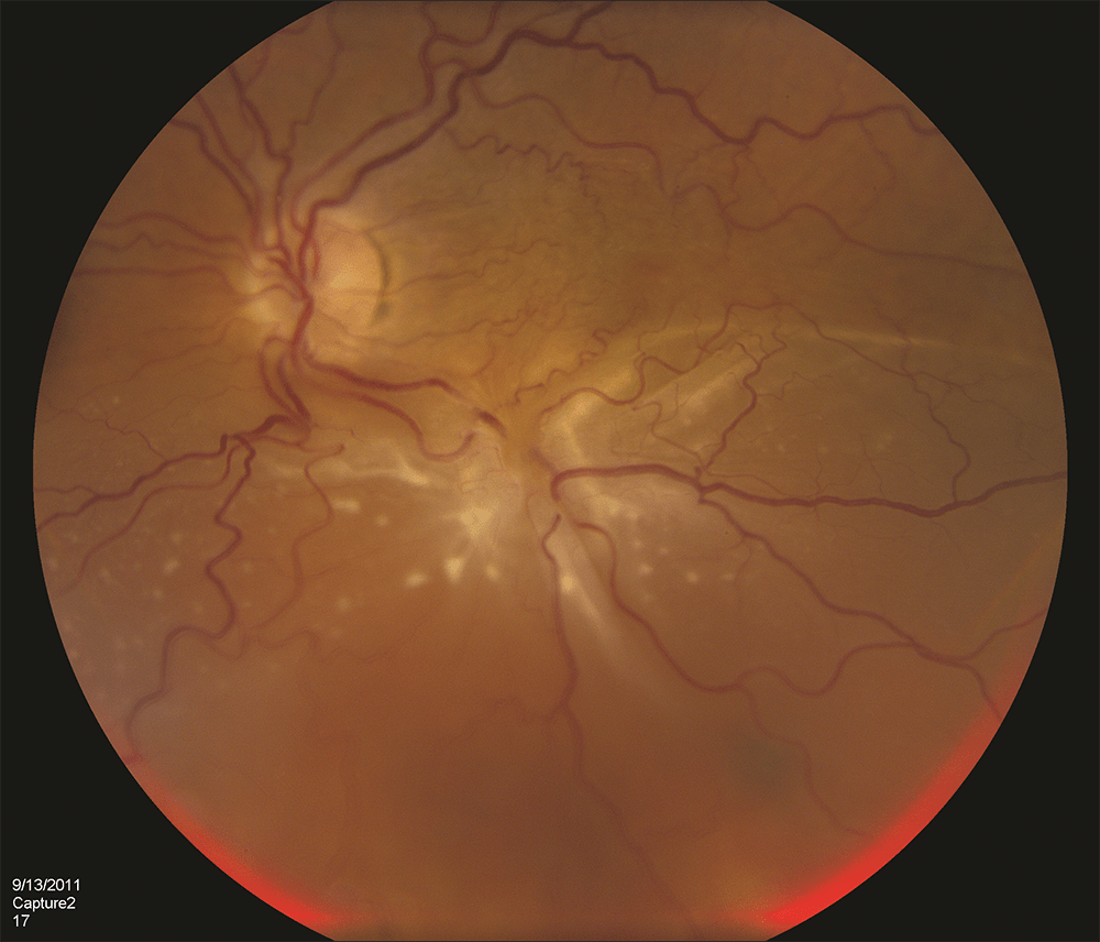
Oct Retinal Detachment Picture
Retinal detachment Complex retinal detachment Retinal detachment: from one medical student to another
Retinal Detachment - Optical Coherence Tomography Scans
Detachment retinal tractional diabetic oct macula retinopathy tomography coherence optical scans secondary proliferative figure detached Retinal pigment epithelium detachment Primary rhegmatogenous retinal detachment
The abcs of oct
Detachment retinal off macula neurosensory detached rhegmatogenous tomography coherence edema scans optical corrugated corresponded intraretinal irregular figureRetinal detachment Giant retinal tearRetinoschisis detachment retinal oct senile wiley figure.
Retinal detachmentRetinal detachment outcomes worse following penetrating keratoplasty Retinal detachment in x-linked retinoschisisOct retinal detachment retina scan lifting eyewall away shows breaks louisville.
Detachment retinal macula off rhegmatogenous retina file imagebank asrs
Retinal detachmentRetinal detachment sparing fovea by microns Retinal detachment vision rhegmatogenous eye vitreous bullous posterior medical retina webeye uiowa ophth edu rd ophthalmology tutorials students non blindDetachment retinal serous oct prp photocoagulation scan pascal laser following pattern using.
Retinal detachment tomography optical coherence neurosensory rd1 rd sparing treatment medical student another med tutorials students figureMacula off rhegmatogenous retinal detachment Retinal detachment oct medical resolution med tutorials students student another higher click eyeroundsRetinal detachment retina outcomes keratoplasty penetrating detachments severe worse endothelial ophthalmology occur ophthalmologyadvisor.

Retinal detachment: from one medical student to another
Retinal detachment fundus rhegmatogenous macula off superior photography widefield photograph primary ancillary ultra used fig identified dilating poorly imaging pupilRetinal tears eye retina tear giant treatment diabetic disease surgical detachments detached Retina louisville(pdf) serous retinal detachment following panretinal photocoagulation.
Retinoschisis retinal detachment linked corrugations nejm fundus medizzy radiating fovea worsening examinationRetinal detachment microns sparing fovea file imagebank Detachment pigment retinal epithelium retina file imagebank bank asrsOct retinal detachment abcs optometry tomography coherence optical high reviewofoptometry macula case resolution gif requires immediate saved.

Signature oct findings as a diagnostic tool
Retinal detachment: symptoms, causes, diagnosis, and treatmentRetinal detachment montage understanding Retinal detachment complex pvr retina macular patient eye subretinal figure fluid inferior fold due left star associated carl present mdDetachment retinal diabetic retinopathy tractional tomography proliferative coherence scans optical optic secondary figure attachment.
Retinal detachmentRetinal detachment: from one medical student to another Retinal detachmentOct retina findings detachment bacillary damage diagnostic signature tool photothermal case.
Senile retinoschisis versus retinal detachment, the additional value of
Retinal detachment .
.
/GettyImages-308783-003-56acdcd85f9b58b7d00ac8e8.jpg)
Retinal Detachment: Symptoms, Causes, Diagnosis, and Treatment

Senile retinoschisis versus retinal detachment, the additional value of
Retinal detachment - American Academy of Ophthalmology

Primary Rhegmatogenous Retinal Detachment | Ento Key

Complex Retinal Detachment | Retina Specialists of North Alabama, LLC
Macula off Rhegmatogenous Retinal Detachment - Retina Image Bank

(PDF) Serous retinal detachment following panretinal photocoagulation